Category Archives: Physical Therapy

Massage therapy can help with TMJ dysfunction

by Nik Panagos, RMT

What is Temporomandibular joint (TMJ) dysfunction?

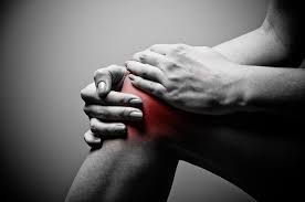
(1) TMJ dysfunction is a disorder of the muscles of mastication (chewing), the temporomandibular joints, and associated structures. This disorder also presents with pain, discomfort, or limitations whilst at rest or movement.

(1) Image above depicts TMJ location and each part of the moving area.

How do I tell if I have issues with my TMJ?

TMJ dysfunction may present with a combination of the following symptoms:

  • Pain in the TMJ at rest or with movement
  • A clicking feel or sound with pain
  • Movements impaired such as chewing/swallowing/opening the mouth/closing the mouth/moving the mouth side to side
  • Headaches
  • Sinus congestion with pain
  • Ear pain
  • Poor posture

I have clicking or popping in my jaw when I chew or yawn, is that normal?
Clicking or popping sound in the jaw is normal as long as no pain occurs or no movements of the jaw are impaired. If none of the listed symptoms occur,then it is not TMJ dysfunction.

Are there different types of TMJ Dysfunction?

The three main types of dysfunctions are:

  1. Internal derangement of the joint: Dislocation, displaced discs, trauma to the lower jaw
  2. Degenerative joint disease: overuse or aging of joint, osteoarthrosis, rheumatoid arthritis, or perforation (puncture) of disc
  3. Myofascial Pain: Muscles surrounding the joint in a hypertensive state (high tension). Usually caused from overuse of clenching of the muscles of mastication (chewing muscles)

What are some common representations of TMJ dysfunction?

(2) Up to 70% of TMJ dysfunction patients suffer from pathology or malposition of the TMJ disc (disc displacement).

Normal movement of the jaw function: the lower jaw opens as the disc slides with the lower portion of the jaw (condyle) allowing for normal opening and closing without pain or dysfunction

Anterior disc displacement with reduction (ADDWR): The disc is displaced slightly forward causing a clicking and potentially some pain as the lower portion of the jaw (condyle) slides over the condyle ending in the proper open position

Anterior disc displacement without reduction (ADDWOR): The disc is displaced so far forward it prevents thejaw from opening fully.

(2) Image above illustrates some common anterior disc displacement of the TMJ

How do TMJ dysfunctions occur?

There are many different factors that can lead to TMJ dysfunction. Some common reasons why it occurs include:

  • Excessive use of mastication (chewing) muscles (I.e. chewing gum, chewing tobacco)
  • Careers with repetitive strain on muscles around the TMJ often due to prolonged seated posture (I.e. call centers, receptionist, instructors)
  • Accidents involving neck/upper back/shoulder trauma (I.e. Moter vehicle accident, contact sports)
  • Dental issues (I.e. missing teeth, uneven overbite or underbite, deviation of jaw)
  • Stress grinding during sleep

How can Massage Therapy help with TMJ dysfunction?

Massage therapy will help with TMJ dysfunction by physically manipulating restricted muscles, ligaments, and joint capsules surrounding the TMJ. Reducing the restricted muscle tissues will allow for movement to resume without pain or limitation. Mobilizing the joint ligaments and stretching will allow for proper function of the joint space to resume. Stretching the joint capsule will reduce pain, improve function, and reduce clicking if present. Manipulation of these structures will also help improve the overall function of the jaw.

What can I do at home to help prevent or reduce symptoms of TMJ dysfunction?

There are many different ways to help with TMJ dysfunction at home; some of the following ways you can help at home are:

  • Self-massage to the muscles of mastication (chewing muscles)
  • Reducing the use of chewing gum or chewing tobacco products
  • Keeping overall dental hygiene in good standing as tooth decay or wear can cause alignment issues that can put extra strain on muscles surrounding the TMJ
  • Follow a prescribed home exercise program to improve the strength and motor control of the muscles that influence the TMJ as well as postural strengthening exercises for the neck and upper back.

Do you treat inside the mouth as well? Is this the only way it can be treated?

Due to the nature of where the TMJ is located, treating within the mouth is usually the best way to access the joint itself for manipulation. However, the TMJ can also be treated without internal mouth manipulation.

In summary, the TMJ is an overlooked area for treatment unless it advances to a point of dysfunction or severe pain. Physical manipulation of TMJ dysfunction is an effective method of treatment for the condition.

Text References

1 Clinical Massage Therapy, Fiona Rattray, Linda Ludwig, Jan, 2005 Pg 597 (Citation 2021-07-13)

2  Meghan K Murphy, BE, Regina F, MacBarb, BS, Mark E. Wong, DDS, and kyracos A. Athanasiou, PhD, PE. November/December 2013. Temporomandibular joint disorders: A review of etiology, Clinical management, and tissues engineering strategies Volume 28, Issue 6 (Citation 2021-07-13)

https://www.ncbi.nlm.nih.gov/pmc/articles/PMC4349514/

Image References

1 Depiction of temporomandibular joint and associating structures, (Citation 2021-07-13), motionspecficrelease.com. https://bodytechphysio.files.wordpress.com/2021/11/5a1f7-4a037c_398c46d6c03c45e0bf0fe91839b2fc33mv2.webp2 A schematic representation of the position of the TMJ disc in three different conditions, (Citation 2021-07-13), researchgate.net.
https://www.researchgate.net/profile/Tarun-Goswami-3/publication/24147487/figure/fig6/AS:668687967195141@1536439043532/A-schematic-representation-of-the-position-of-the-TMJ-disc-in-three-different-conditions.png

Management of Osteoarthritis

What is Osteoarthritis (OA)?

Osteoarthritis is the most common form of arthritis and occurs when the protective cartilage that provides cushion and support at the ends of bones gradually wears down. This is a degenerative disease that can worsen over time, often resulting in chronic pain affecting your day-to-day activities. Eventually if the cartilage wears down completely, the bones in the joint will rub directly on each other exacerbating the symptoms.

Common Symptoms of OA

  1. Pain and Tenderness
  2. Joint Stiffness
  3. Muscle Weakness and Loss of Flexibility
  4. Grating Sensation
  5. Bone Spurs- not a symptom but a sign
  6. Swelling

Most Common Risk Factors

  1. Old Age
    • Articular surfaces on the end of bones can wear down over time due to the natural aging process that occurs to muscles, joints, and bones.
  2. Obesity
    • Excess weight puts more stress on the joints (commonly occurs to weight bearing joints such as the hip and knee).
  3. Repeated Stress on the Joint
    • Over time this will cause the articular surfaces to wear down.
  4. Joint Injuries
    • A break or tear can lead to the development of OA over time.
  5. Genetics and Certain Metabolic Diseases
    • People with a family history of OA are at a greater risk.

Treatment for OA

An effective approach is seeking physiotherapy treatment at earlier stages, accompanied with an exercise program specific to you. You may feel some discomfort during exercising, but this feeling is normal and should calm down. If it is unbearable then do not continue with the exercise. Of course with any exercise, there are always risks associated, therefore consult your doctor prior to beginning any new exercise program.

                Exercise has many benefits for ALL people including improved health, fitness, and mood. Many people believe that exercising with OA could harm your joints and cause more pain, but research shows that people can and should exercise when they have OA. It is considered the most effective, non-drug treatment for reducing pain and improving movement in those with OA.

Recommended Types of Exercise

So now we know that exercise can ease symptoms of arthritis, but what is best for you? Well that varies from person to person so here are a few main categories of exercises to include in your program:

  1. Flexibility

                This category includes exercises that will help to improve or maintain the range of motion of the affected joint(s). By relieving stiffness in the joint and increasing the ability for the joint to move through its full range, you will decrease the risk of further damage, improve the function of the limb and joint, and decrease overall pain. There are two main categories of stretching to consider:

  • Dynamic Stretching: these are movement-based stretches that involve multiple joints. They should be performed prior to activity to prepare the body.
  • Static Stretching: these are stretches that take the muscle to its end range before holding that position for a minimum of 30 seconds. These stretches should be performed after activity when the muscle is already warmed up.

2. Strengthening

                These exercises work to build stronger muscles to help support and protect the joints. This allows for offloading of the affected joint which has the potential to relieve many symptoms. It is recommended to engage in strengthening exercises 2-3 days/week. Examples of strengthening exercises include lifting a limb against gravity, using free weights or elastic bands, or weight machines requiring you to push or pull against resistance.

3. Aerobic / Endurance

                These exercises help with overall fitness and improve your cardiovascular health. They typically involve the use of large muscle groups in the body in a repetitive and rhythmic manner. Canada’s guidelines for adults are to achieve 150 minutes of moderate- to vigorous-intensity aerobic physical activity per week. This intensity typically involves you to breathe a little harder and sweat but not be out of breath. Activities falling under this category include walking, biking, dancing, or even everyday activities such as mowing the lawn or shoveling as long as you are achieving a moderate- to vigorous-intensity.

4. Balance

                This is a fourth, less recognized category that is very important to consider in your exercise plan. These exercises will target smaller groups of muscles to decrease your risk of falling and can help improve your ability to do other exercises. Balance exercises include anything with a smaller or unstable base of support such as performing activities on a foam pad or single legged exercises.

Summary

                Arthritis doesn’t have to keep you from living your life and participating in your everyday activities. Exercise and arthritis should coexist! Research has shown that people with osteoarthritis who exercise regularly have less pain, more energy, improved sleep and a better day-to-day function. Work with your Physiotherapist to create an exercise program that is right for you and kick start your road to a healthier life!

BodyTech Physiotherapy

Why is your core so important?

By Courtney White, Registered Physiotherapist

Have you ever wondered what makes up your core, why is it important, or how to safely train your core without getting back pain? Keep on reading to find out these answers and more!

Your core is more than just the muscles that you can see. There is a group of muscles below the big six pack muscles that wrap around you like a corset. Their job is to support you during every movement. This inner unit is like a TRANSFER STATION. To get power generated from your legs all the way to your shoulders, it must pass through the inner core. So, if you do not have proper control over your core, that power will not move between your upper and lower body as smoothly as you want it to.

What Makes Up the Inner Core Unit?

The inner core consists of the following: the diaphragm, the pelvic floor, transversus abdominus (TA), and multifidus. Collectively, these 4 structures create a CORE CANISTER.IMG_5235-01Pic for Core

Diaphragm: The diaphragm is our primary breathing muscle and it forms the top of the core canister. The diaphragm is the component that has the primary control over the intraabdominal pressure within the canister. It moves up and down as you breathe which changes the space in the abdomen and as a result, influences the pressure within the core canister.

Pelvic Floor: The pelvic floor is a group of muscles that form the bottom of the canister. It supports the weight of all the internal organs within our abdomen, assists with bowel and bladder control, and helps to control the intrabdominal pressure along with the diaphragm. The pelvic floor is partners with the diaphragm. When the diaphragm moves up and down, so should the pelvic floor.

Transversus Abdominis (TA): This muscle connects the top and bottom of the canister as well as creates the front and sides of the core canister. It runs deeper than the external six pack muscles that you can see. Its role is to support your lower back during movement and transmit forces between your upper and lower body.

Multifidus: This muscle forms the back of the core canister and runs along the spine. Multifidus serves as a primary support for the spine, pelvis, and hips.

[1]Canister

What Are the Functions of the Core Canister? 

  1. To support the back, pelvis, hips, and trunk during movement
  2. To serve as a transfer station for power and energy between the upper and lower body
  3. To assist with bowel and bladder control
  4. To improve performance during physical activity

How Does the Core Canister Work?

To gain a stronger core overall, it is important to learn how to “pressurize” the core canister. Previously, inner core training has focused largely on tensing the walls of the core canister through focused contractions of TA. Learning how to activate the diaphragm and pelvic floor was often missed or brushed over. More recent evidence now suggests that breath is the driving force behind influencing the intrabdominal pressure. Therefore, the diaphragm is the leader behind pressurizing the core.

When you breathe in, your diaphragm contracts and pushes down into the abdominal cavity which increases the intraabdominal pressure. In response to this increased pressure, the pelvic floor relaxes and lengthens to accommodate the abdominal organs translating downward. During your inhale, the pressure in the front part of the core canister is increased as your belly expands. This is counteracted by multifidus in the back as well as TA which work together to try and keep the intraabdominal pressure evenly distributed. When you breathe out, the diaphragm relaxes and rises which decreases the pressure within the core cannister. As a result, the pelvic floor should normally contract and rise.

[2]GifCore blog

How Do We Train the Core Canister? 

Many people focus on crunches, planks, and other large abdominal exercises when training the core but negate breathing. Learning to connect your breath with movements is the key to developing a stronger core. It will allow you to control the intrabdominal pressure within the core canister. Exercises like planks and crunches are not necessarily bad exercises as they can be very effective later if you first learn to optimize the control of your core canister.

The first step is to learn how to engage your diaphragm through diaphragmatic umbrella breathing. Once you have achieved this, it is time to add in the pelvic floor so that you learn how to engage your diaphragm and pelvic floor together as a unit, referred to as piston breathing. You can also learn how to connect and activate TA and multifidus to further optimize your control over the core canister. However, it is important to highlight that the goal here is to learn how to change the size of your core canister by recruiting all these muscles together, rather than focusing solely on how to contract each specific muscle. Once you have learned how to control the pressure within the core canister, it is now time to perform bigger movements that require you to maintain control over the canister while doing a larger activity.

Check out our post on our Facebook and Instagram pages (dated May 21, 2020) for exercises you can try at home to increase your control of the core canister!

What Can Happen If You Do Not Have Control Over Your Core? 

It is not uncommon to have a weak inner core or lack control over your core canister. This can originate from prolonged sitting and slouching which distorts and changes the shape of the core canister (see part B in Figure 3 below). This positioning leads to breathing through the accessory muscles in your neck and chest, decreasing the engagement of your diaphragm. As you can see in the figure below, this puts a lot of strain on the lumbar spine which can lead to pain and injury. Ideally, your core canister should look something like part A in the figure below where the intraabdominal pressure is equally distributed. Without proper control over your inner core, it makes it very difficult for you to be able to hold your body in a good position during exercise and physical activity. This increases your risk for injury.

[3]DNS-Scissor

Now that you have learned more about what the core is and why it is important, if you have any questions or are interested in setting up an appointment with one of our Physiotherapists, contact us today! We will be happy to help you achieve your goals by developing an individualized treatment plan. 

BodyTech Physiotherapy

Text References

Diane Lee & Associates: core training vs. strengthening (internet). South Surrey: D G Lee Physical Therapist Corp; (cited 2020 May 22). Available from: https://dianeleephysio.com/education/core-training-vs-strengthening/

Key J. ‘The core’: understanding it, and retraining its dysfunction. Journal of bodywork and movement therapies. 2013 Oct 1;17(4):541-59.

Pronatal Fitness: the first move to teach your clients (internet). 2018 July 18. (cited 2020 May 22). Available from: https://pronatalfitness.com/2018/07/18/360-breathing/

Image References 

[1] The role of the diaphragm. Digital Image. Chiroup.com. Nov 2016. [Accessed on 2020 May 22]. Available from: https://chiroup.com/the-role-of-the-diaphragm/. 

[2] Breathing variations. Digital Image. S. McLaughlin. Aug 2019. [Accessed on 2020 May 22]. Available from: https://www.alignforhealth.com/self-care-for-pain/category/core%20stabilization 

[3] The “pop can” core. Digital Image. J Smeaton. Apr 2019. [Accessed 2020 May 22]. Available from: https://www.depthtraining.ca/the-pop-can-core/.

Is Virtual Physiotherapy for you?

knee painBodyTech Physiotherapy is offering virtual physiotherapy appointments with our most experienced therapists. All you need is a phone, tablet or computer. It is as simple as clicking a secure link to connect with us in a video chat.

What to expect?

Your first appointment with us will be an assessment. The Registered Physiotherapist will ask questions to complete a history and gain a complete understanding of your injury in order to provide a diagnosis.

You will be asked to do a series of movements to allow the therapist to examine your range of motion and strength, as well as be able observe how you move and perform certain tasks. You may be asked to demonstrate walking, stairs, sitting, standing or any movement pattern specific to your injury.

Treatment

Based on the findings from your assessment, the therapist will be able to cue and correct your movement patterns. You will also be given specific exercises to help in your recovery. Your therapist may give you some self-directed treatment techniques, or teach another person how to assist you with these techniques.

You will have a chance to ask questions and clarify your abilities and restrictions. Your therapist will educate you about your condition and provide suggestions on how to modify your activities if needed.

Still not sure if virtual physio is right for you? Schedule a free 10 min phone consult with one of our therapists.

Call 519-954-6000 or email: info@bodytechphysio.ca

BodyTech Physiotherapy

Concussion Management Part 2: How Long Does Recovery Take?

By Cassandra Kroner, PT

In part 1 of the concussion management blog series we covered how the brain is affected following a concussion, common symptoms, why early intervention is critical, and how physiotherapy can help optimize recovery. One of the most frequent questions people have following a concussion is about recovery time – ‘when can I go back to work full time?’ or ‘when can my son/daughter play soccer again?’. It can be helpful to understand the general stages of injury and potential progression of symptoms:

concussion blog image_jan2019

The initial days following a concussion are considered the acute stage of injury, and cognitive and physical rest is critical at this time. After 7-10 days of adequate rest the chemical balance and blood flow in the brain has been restored, and symptoms that continue are known as post-concussion syndrome. Some symptoms can last upwards of 6 months or years post injury. It is important to keep in mind that not everyone will progress through all three stages, and the length of time symptoms last will vary between individuals.

Why Recovery Can Take Longer

There are a number of factors that can complicate and prolong recovery, and these can help us predict if symptoms are likely to persist longer than the usual 6 weeks. A history of migraines, mental health conditions such as depression or anxiety, or learning disabilities, have been found to increase recovery times. Additionally, visual or vestibular dysfunction or a high number of initial symptoms following a concussion usually indicate prolonged recovery.

History of Concussion

People who have had a previous concussion are more susceptible to have another one due to a lower threshold for injury after each concussion – meaning the next concussion can happen from a lower severity injury than the first time. Additionally, there is often an increased number of symptoms and a longer recovery time after each subsequent concussion. A concussion at a young age risks disruption of brain circuits yet to be developed, and also creates a wider window for repeated future concussions.

Repetitive hits that are common in sports such as hockey or football, which do not cause a concussion, are known as sub concussive trauma. Research has shown this repetitive trauma can result in increased reaction and processing time, memory impairments and increased chance of making mistakes. These effects can place an athlete at an increased risk for a concussion during sport. If the athlete does sustain a concussion at this point, the brain has a diminished reserve capacity to manage injury, and the effects of subsequent concussions are cumulative and result in increased impairment in function with each concussion.

Additional Injuries

Another complicating factor is the presence of other injuries, such as whiplash or neck sprain/strains, that can occur with falls or car accidents. These neck injuries alone can cause similar symptoms to a concussion including headaches and dizziness, and in combination with a concussion can result in more severe and prolonged symptoms. Having an assessment by a physiotherapist can determine which symptoms are from the neck injury and which are from the concussion – resulting in individualized treatment strategies to target the cause of each symptom.

To conclude, although concussions can be an invisible injury, they need to be properly managed and rehabilitated just like any other injury. This management includes assessment to determine the cause of symptoms, specific treatments to address each impairment, and strategies to manage recovery at home. Visiting a physiotherapist trained in concussion rehabilitation will ensure that both concussion symptoms and neck injuries are addressed. The goals of treatment are to restore physical and cognitive function while facilitating a safe return to work and sport. Awareness and education about concussions and treatment options are important to ensure that people don’t suffer unnecessarily from prolonged symptoms – this is where a trained Physiotherapist can help!

BodyTech Physiotherapy

Osteoporosis and Exercise

By Lauren Harding, Registered Kinesiologist

What is osteopenia and osteoporosis?

unnamedOsteopenia and osteoporosis are conditions characterized by a loss of bone mineral density (BMD). BMD is a measure of the quantity of minerals (calcium and phosphorus) in a precise volume of bone. The difference comes in their severity.

Osteopenia indicates a lower BMD causing bones to be generally weaker. This bone weakness becomes more pronounced with osteoporosis as the bones become more brittle and porous (filled with air pockets), ultimately increasing the risk of fracture. Though less severe, osteopenia is a serious risk factor for developing osteoporosis.

Osteoporosis is often labeled as the ‘silent thief’ since bone loss occurs gradually, typically without symptoms. However, these four factors can signal underlying osteoporosis:

  • Loss of height over time and/or development of a stooped posture
  • Sudden back pain without any obvious cause
  • Fracturing after a seemingly minor incident


How are these three conditions treated?

An excellent approach is early intervention physiotherapy treatment, accompanied by a well-designed exercise program. It is important to consult your doctor before starting a new exercise plan. As with any exercise, there is always risk associated.

Osteoporosis Canada recognizes exercise not only for increasing cardiovascular endurance, but also as a fundamental component to protecting your bones. Exercise maintains bone mass and builds muscle strength, as well as increases flexibility and range of motion, balance and coordination. Benefits also include reduced pain and inflammation, while promoting loss of excessive weight. Additionally, the risk of falling is reduced.


What types of exercise should I be doing?

There are 5 types of exercises recommended for individuals with osteopenia and osteoporosis:

Types of Exercise How Often Should I Do These?
Aerobic 3 to 5 days per week, a minimum of 150 minutes per week
Strength Training 2 to 3 times per week
Balance Every day
Stretching Every day
Posture Training Always


Aerobic Exerciseunnamed

Aerobic exercise is considered any continuous, rhythmic activity that strengthens and stimulates the heart and lungs, thereby improving the body’s use of oxygen. It is recommended that ALL adults get 150 minutes of moderate to vigorous physical activity a week, or about 20 to 30 minutes per day, for at least 10 minutes at a time. During moderate-intensity activity, your heart beats faster and you breathe harder. The rule of thumb is that you are able to talk during these activities, but you are working too hard to be able to sing. During vigorous-intensity activity, your heart beats faster still and you continue to breathe harder. You are unable to say more than a few words at a time during vigorous activity. It is important with osteopenia and osteoporosis to do weight bearing aerobic exercises. Examples of weight bearing aerobic exercises include:

  • Brisk walking, dancing, stair climbing, running, step aerobics, hiking, jogging, jump rope, and treadmill walking/running


Strength Training

Elderly_exercise

In order to increase your muscular strength, you must work against resistance until your muscles feel tired.

This helps to reverse muscle atrophy, a condition that occurs when your muscles start to waste away due to lack of use. Furthermore, resistance training strengthens the muscles surrounding your joints, ultimately reducing further joint damage and decreasing risk of injury. ALL adults should do 2-3 days of strength training a week. Exercises using free weights (dumbbells), exercise bands or weight machines are strongly recommended. Examples include:

  • bicep curls, sit-to-stand, lunges, rows, calf raises, bridges, triceps kickbacks, countertop push ups


Balance Exercises

Balance exercises help maintain your footing when an unexpected movement occurs in your daily life. Improving balance and coordination can reduce your likelihood of falling, therefore decreasing your fracture risk. Ironically, in challenging your balance, you run the risk of falling. For this reason, you must always take precautions such as having a chair nearby to hold onto. When you are training your balance, there are two main types of balance exercises:

  • Static Exercises – Stand still in one spot holding a certain posture in order to practice balance. For example, standing on one foot on the floor.
  • Dynamic Exercises – Balance is challenged more with adding movement. For example, walking ina straight line while touching the heel of one foot to the toe of the other with each step.


Stretching

unnamed (1)As you age, you lose flexibility which can increase your stiffness and discomfort, often preventing you from staying active. Stretching exercises help you to counteract this by increasing the range of motion of your joints and improving your flexibility. It is important to note that stretching should always be done after the muscles and the body are warmed up since stretching cold, stiff muscles increases your risk of injury. Just like in balance training, there are two main ways to stretch:

  • Static Stretching – Take the muscles to their end range of motion and maintain that position for at least 30 seconds in order to enhance soft tissue and muscular flexibility.
  • Dynamic Stretching – Functional, multi-joint movements that typically increase in range of motion and speed as the body begins to warm up.

While both are effective, dynamic stretching has been shown to be more beneficial than static stretching as a way to warm up prior to activity since it has been found to improve balance, strength, reaction time and agility. Static stretching, however, is still important and is most beneficial when performed at the end of exercise as a cool down. Static stretching can also be done separately when the body is warm as part of an everyday attempt to improve body mechanics, posture and flexibility.


Posture Training

unnamed (1)We all have a natural curve in our spine, however, weak back muscles and/or spinal fractures can cause an excessive forward curvature of the spine. Rounding of the upper back is known as exaggerated kyphosis. This puts pressure on the front of your vertebrae, placing them at even great risk of fracture. Posture training exercises help to improve the alignment of your spine by correcting shoulder, back and neck positioning. Focus should be placed on exercises that strengthen the back muscles and reduce forward head posture. Abdominal exercises that strengthen the core muscles, help to maintain good posture as well.


To Sum Up

An exercise program is a vital component in the management of osteoporosis.

Regular participation in aerobic and strength training is fundamental, as well as balance, posture management and stretching.  All in all, physical activity for individuals with osteopenia or osteoporosis can promote bone health and overall quality of life.

BodyTech Physiotherapy

Concussion Management Part 1: The Role of Physiotherapy

By Cassandra Kroner, PT

What is a Concussion?

Concussions are a type of mild traumatic brain injury. Common causes include car accidents, sports, falls, or workplace accidents. Concussions can result from direct impact to the head, or from forces elsewhere in the body such as sudden acceleration or deceleration that cause an  injury to the brain and brain-stem.  The result is damage to cells and chemical imbalances that disrupt normal brain function.

Concussion Head imageImmediately following injury a sequence of chemical processes occur as the brain attempts to restore its normal balanced state. This increased activity in the brain is happening at a time when blood flow is decreased to the site of injury, creating an increased demand for energy. The resulting impairments in neurological function can cause a variety of signs and symptoms:

Physical Behavioural/Emotional Cognitive
Headache Drowsiness or fatigue Feeling foggy
Nausea Irritability Trouble thinking clearly
Vomiting Depression Feeling slowed down
Blurred or double vision Anxiety Difficulty concentrating
Balance problems Sleeping more than usual Difficulty remembering
Dizziness Difficulty falling asleep Trouble finding words
Sensitivity to light or noise Sadness Confusion


First Steps Following Injury

Concussions are often under-reported and misdiagnosed, and it is important to note that loss of consciousness is not necessary for a diagnosis. Contributing to the difficulty in identifying concussions is the lack of imaging or other tests to aid in diagnosis. Unless there is bleeding or swelling in the brain, the changes that occur with a concussion are not visible on a CT or MRI. If a concussion is suspected, an evaluation by a physician is recommended, and unless symptoms are severe or quickly worsening it is usually not necessary to visit the emergency room. Once the diagnosis is established and conditions requiring further medical treatment are ruled out, treatment should begin immediately.

Early Management

HeadacheTimely intervention following a concussion is essential to ensure optimal management and recovery. An outdated approach to concussion treatment is to stay in a quiet dark room until symptoms are resolved. With a growing demand for evidence-based treatment strategies, there is a wealth of new research that refutes this old-fashioned ‘dark room’ approach. Although complete rest is recommended for the first 48-72 hours after injury, research supports a more active approach to recovery following the initial rest period. Prolonged physical rest can lead to de-conditioning, depression and fatigue, making it more difficult to return to the previous level of physical activity.

Complete physical and cognitive rest immediately following a concussion is critical to ensure adequate energy supplies for the brain as it attempts to heal. Excess physical or cognitive exertion at this time will use precious energy that the brain needs and can result in exacerbation of symptoms and prolonged recovery. Physical rest means no exercising and caution with exertion around the house. Cognitive rest should focus on refraining from activities that require concentration (schoolwork, reading), as well as visual attention (television, video games, computer or phone use). Alternative options are listening to music or audio books.

Importance of Physiotherapy

A visit to a physiotherapist with advanced concussion management training is recommended for a detailed assessment following a concussion. Your physiotherapist will take a thorough history and can assess visual and vestibular symptoms, balance, cognitive function, and any additional injuries sustained at the time of concussion. Recommendations for the initial rest period as described above will be tailored to each individual, and further suggestions for management of symptoms will be provided.

Treatment plans involve a carefully monitored graded program of exertion to assist with a safe return to work/school and then sport. Every individual will experience a different set of symptoms following concussion, and as a result there is not a ‘one size fits all’ approach to treatment. This is why having a professional guide you through recovery is valuable. Specific and progressive exercises will be provided to target deficiencies in the vestibular and visual systems. To facilitate a gradual return to school or work, suggested accommodations would be provided to minimize symptoms and maximize participation. Additionally, manual therapy to address complaints such as neck pain or headaches can be part of treatment. Once the individual has returned to school or work, physical exertion testing is the last step before being cleared for sport.

Recovery time frames vary between individuals, but for many people, symptoms resolve in a month or less. However, there are a number of factors that can delay or prolong recovery. Stay tuned for part 2 of the concussion management blog series to learn more.

BodyTech Physiotherapy

Update: Continue reading on part two of our Concussion Management blog series.

Physiotherapy after a Fracture

By Courtney Lacey, PT

fractureIf you have recently broken a bone, you may be wondering when you will be able to return to all of your normal activities. While it typically takes 4-8 weeks for a bone to heal, you will likely require physiotherapy to help get you back to full function.

How do fractures happen?

A broken bone, also known as a fracture, can occur in many ways. Most often, broken bones are the result of a traumatic mechanism of injury such as a fall, motor vehicle accident or contact during a sporting event. Fractures can also occur from repetitive motions which place stress on the muscles and bones. A common example of this is stress fractures in the legs from running. Finally, fractures can more easily occur in people with osteoporosis – a disease which weakens bones and makes them more likely to break.

How do you know if you have a fracture?

These are some signs and symptoms which may indicate that you have a fracture:

  • Immediate and severe pain following a fall or accident
  • A “pop” or “click” heard or felt during the incident
  • Swelling in the area
  • A bump or deformity
  • Unable to weight-bear through the injured limb

If you suspect you have a fracture, you will need to see a doctor who will order an X-ray to confirm the diagnosis. Often, those who experience an injury causing a fracture will go to the hospital to be evaluated.

Does a fracture heal?

While bone healing takes approximately 4-8 weeks, the timeline depends on both the person and the type of fracture.  In order for a bone to heal properly, it has to first be set in the proper position, which is called reduction. The doctor may be able to reposition the bones without surgery, which is called a closed reduction. Sometimes, surgery may be required to bring the ends of the bone close together, which is called an open reduction. Pins, plates or screws may also be used to keep the bones in place. If the fracture did not cause any part of the bone to shift out of place, no reduction is needed. Once the doctor has determined the bones are in a good position to allow for healing, the area will be immobilized in a cast or a splint.

When can the cast come off?

To determine if you are ready to have the cast removed, you will have an X-ray done with the cast or splint in place. The doctor will look for the formation of a callus, which demonstrates that healing has taken place. The doctor will then remove the cast and may recommend that you have physiotherapy. Physiotherapists play a key role in returning you to your full function as quickly as possible after a fracture.

Why do I need physiotherapy?

There are several reasons why physiotherapy is needed after fracture. Depending on the amount of healing that has occurred, your doctor may have special instructions (how much weight to put through the limb, certain activities to avoid, etc.) that your physiotherapist can help you understand. Once the cast is removed, you may still have some swelling and pain around the fracture site. Physiotherapists may use modalities (such as ultrasound or TENS) to help decrease pain and swelling and improve your mobility and tolerance for using the injured limb in daily activities. If you had surgery, you may also have a scar which creates scar tissue and can disrupt movement. At BodyTech Physiotherapy your therapist will use manual therapy techniques to help mobilize the scar tissue and the areas around the injury as needed to  restore normal movement around the surgical site.

Physiotherapy is crucial to improve your functional mobility that you may have lost during your time in the splint or cast. Immobilization over 6-8 weeks will cause loss of range of motion and strength, which will make daily tasks difficult to do. Your physiotherapist will help restore your proper range of motion using manual therapy techniques. While the fracture site will be stiff and sore, you may also lose range of motion at surrounding joints that were moving differently during the healing process. For example, if you have broken your elbow, it is also necessary to  assess your shoulder, wrist and hand to ensure that these joints are moving properly. Not correcting the mobility around the fracture site can prolong your healing process and lead to future injuries as well.

Once your range of motion has been restored, you will need to regain strength in order to return to your pre-injury activities. Your physiotherapist will work with you to create a proper strengthening program to re-introduce your bones to loads and stresses that you encounter in your daily activities. Lack of strength or going back to activity too soon puts you at risk of re-injury or prolonging the healing process. Physiotherapy will help you understand the correct exercises to do and will tailor your program to the activities you plan to return to, whether it be high level sport or recreational activity.

How long until I am back to my regular activities?

Your rehab program will vary in length depending on the type of fracture, if there was surgical intervention, and the type of activity you plan to return to. Depending on the nature of the injury, physiotherapy can take anywhere from 8 weeks to one year for more complex fractures. Your physiotherapist will guide you through your rehab program, ensuring you are progressing at an appropriate rate and prevent complications or future injury.

BodyTech Physiotherapy

Frozen Shoulder/Adhesive Capsulitis

By Carla Cranbury, PT

What is it?

Frozen shoulder, also called adhesive capsulitis, is a gradual onset shoulder condition characterized by pain and limited range of motion. This is caused by inflammation and tightening of the shoulder capsule. Typical initial symptoms are pain midway between the shoulder and the elbow and difficulty reaching behind the back. Most women will report that they have difficulty doing up their bra and men difficulty putting on their belt.

Why does it happen?

Limited research has been able to discern one certain cause of frozen shoulder – in short, we don’t know. We do know that it is most common in middle aged women (aged 40-65) and people with diabetes. It also is more likely to occur after a virus, a lingering shoulder injury or after shoulder or upper limb surgery.

How long does it take?

Frozen shoulder goes through three main stages, each of which can take weeks to months:

  • Freezing – pain is noticed and range of motion becomes progressively limited
  • Frozen – pain is reduced, but range of motion is further restricted
  • Thawing – pain is reduced and range of motion gradually returns

Can physio help?

Physiotherapy cannot speed up the course of the condition – everyone has to go through each of the three stages in order to recover. The total process of frozen shoulder can take one to two years to resolve.

What physio can do is help you retain function while going through frozen shoulder, decrease some pain, and ensure a full recovery. Maintaining mobility through the process is important and is where physiotherapy can help the most. Physio will also help prevent other injuries that can be caused by compensating for the frozen shoulder – this is especially significant as it is common for the other shoulder to get the same condition.

Your physiotherapist will give you exercises to maintain as much movement as possible and instruct you on how to perform them properly to ensure you are not compensating for the limited range of motion. Hands on manual therapy will help stretch out the capsule to make the exercises easier to perform. Modalities such as ice, heat, TENS, and acupuncture can also be used to decrease pain.

Though frozen shoulder can be a lengthy and frustrating process, the right care can make it more manageable and prevent any further complications.

BodyTech Physiotherapy


Reeves B. The natural history of the frozen shoulder syndrome. Scand J Rheumatol. 1975;4:193–6.[PubMed]
Greene WB. Essentials of musculoskeletal care. 2. Rosemont, IL: American Academy of Orthopedic Surgeons; 2001
Pal B, Anderson J, Dick WC, Griffiths ID. Limitation of joint mobility and shoulder capsulitis in insulin- and non-insulin dependent diabetes mellitus. Br J Rheumatol. 1986;25:147–51. doi: 10.1093/rheumatology/25.2.147. [PubMed] [Cross Ref]
Bridgman JF. Periarthrits of the shoulder in diabetes mellitus. Ann Rheum Dis. 1972;74:738–46.
Hazleman BL. Frozen shoulder. In: Rockwood CA Jr, Matsen FA III, editors. The shoulder. 2. WB Saunders: Philadelphia; 1990.
Harryman DT, Lazurus MD, Rozencwaig R. The stiff shoulder. In: Rockwood Cam Matsen FA, Wirth MA, Lippitt SB, editors. The shoulder. 3. Saunders: Philadephia; 2004.

Injury Prevention and Physiotherapy

By Cassandra Kroner, PT

best-chicago-group-step-class.jpgIt is a common misconception that you only need to see a physiotherapist if you have an injury or pain. Physiotherapists have a wide range of skills, and recognizing the risk for future injury is one of them. Injury prevention is applicable to all individuals, regardless of their activity level, from the office worker to the athlete, and especially for those with previous injuries that could reoccur.

Repetitive Strain Factors

  • Occupation
  • Training errors
  • Age
  • Excessive or repetitive loads
  • Inappropriate footwear
  • Structural abnormalities
  • Muscle control
  • Core stability
  • Joint alignment
  • Muscle strength and flexibility imbalances
  • Previous injury
  • Posture

In general there are two types of injuries – traumatic and repetitive strain. Traumatic injuries are easier to understand, as they are the result of a singular event that causes damage. On the other hand, repetitive strain injuries occur when stressors that normally do not cause harm are repeated to the point of causing micro trauma that builds over time until the tissue becomes inflamed and injured. These injuries tend to begin subtly, and gradually increase in severity. Combined with the multi-factorial list of possible contributing factors, repetitive strain injuries can be challenging to diagnose and treat. However, in most cases repetitive strain injuries can be avoided with a good injury prevention and maintenance program.

A Common Factor: Muscle Imbalance

When an individual is involved in one specific sport or has a repetitive aspect to their job, the muscles are put under a great deal of strain to repeatedly perform the same movements. Sedentary jobs involving prolonged postures can have similar effects with certain muscles constantly working for long periods. Over time, muscle imbalances develop as the muscles that are being used the most continue to get strong while the reciprocating muscles become lengthened and weak. These muscle imbalances can cause movement restrictions that affect performance and increase stress on the body. Increased stress on muscles, joints and ligaments eventually leads to a repetitive strain injury.

15250-a-young-woman-stretching-outdoors-before-exercising-pv-630x390.jpgThe First Steps Toward Injury Prevention

Getting started on the road to injury prevention is as simple as booking an appointment with a physiotherapist. If you are unsure how injury prevention could apply to you or if you could be at risk for injury, speaking with a physiotherapist can help clarify your needs and goals. The same expert assessment skills physiotherapists use to diagnose injury will be used to proactively assess for risk factors that could lead to future injury. Your lifestyle, the demands of your job, and the specific sports or activities you participate in will be considered, along with the assessment findings, to develop an individualized treatment plan. This plan could involve manual therapy to correct joint restrictions or muscle length imbalance, strength and flexibility exercises, correction of movement patterns, and education.

By addressing several predisposing factors to injury, the body is optimally prepared to handle the demands of the workplace or sport. This can translate to decreased frequency and severity of future injuries, with the added benefit of a shortened recovery time in the event an injury does occur. Don’t wait for an injury to strike to take charge of your health!

BodyTech Physiotherapy